|
XI.
Chagas Disease
|
|
Case 61:
Chagas Disease / Dermatophytosis
|
|
|
|
Chagas Disease
|
|
Dermatophytosis
|
|
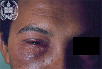
Fig.61-A1
Chagas Disease
The primary infection takes place by the sting of the vector, in Venezuela called Rhodnius prolixus, with the production of a chagoma in the skin, as described in previus case, or of the socalled Romaña sign, i. e.a blepharitis, we are presenting in this case. It is one-sided.
|
|

Fig.61-B1
Dermatophytosis
In this case a fungal infection provoked a blepharitis.
|
|

Fig.61-A2
Chagas Disease
The parasite producing the infection reaching the bloodstream is the protozoan Trypanosoma cruzi with his typical flagellum.
|
|
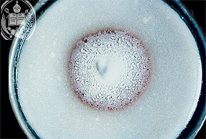
Fig.61-B2
Dermatophytosis
Trichophyton rubrum was the causing agent. Here we show a culture colony of this fungus.
|
|

Fig.61-A3
Chagas Disease
The habitat of the vectors, the Triatomidae, harbouring the trypanosomes, in its found mostly in the roofs of the huts (ranchos) in our country.
|
|

Fig.61-B3
Dermatophytosis
Microscopically T. rubrum is seen in this smear of the culture.
|
|

Fig.61-A4
Chagas Disease
Here we show the Triatomidae called Rhodnius prolixus in different sizes with their eggs.
|
|
|
|

Fig.61-A5
Chagas Disease
In this infestation mostly the heart and the myocardium are affected with the production of a typical acute myocarditis accompanied by a marked hypertrophy and dilatation of the ventricles, as shown in this picture.
|
|
|
| español | english | deutsch |
|
|