|
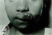
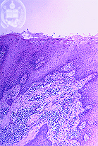
Fig.6-A1
Chronic mucocutaneous Leishmaniasis
This patient from Barinas / Venezuela showed several painful ulcers covered by crusts on the lips, mainly on the upper lip and the palate. This disease began one year ago.
|
|
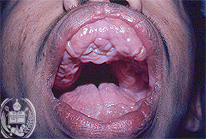
Fig.6-B1
Hyperplastic gingivitis
The gingiva shows a tumor-like proliferation, especially of the superior parts. This tumor tissue is firm and has a whitish to pink color.
|
|
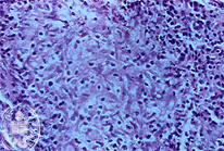
Fig.6-A2
Chronic mucocutaneous Leishmaniasis
Histologically a non-specific granulation tissue with foci of epitheloid cells is found. Parasites are difficult to detect and must be searched for by a higher magnification. The Grocott stain is negative.
|
|
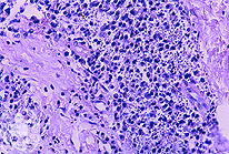
Fig.6-B2
Hyperplastic gingivitis
In the granulation tissue occasionally granular elements are seen representing nuclear detritus. They should not be confused with leishmanias. The Grocott stain is negative.
|