|

Fig.5-A1
Chronic mucocutaneous Leishmaniasis
A patient from the State Apure of Venezuela, a tropical flat area of this country, came to the physician because he suffered since more than one year a chronic inflammatory process in his oral cavity. Now he can hardly open his mouth. There exist extensive ulcerations of the oral and nasal mucosa.
|
|

Fig.5-B1
Carcinoma
In the mucosa of the oral cavity of this 64-years old patient from Barinas / Venezuela extensive necrotic areas are found. Furthermore, a skin nodule at the chin is present.
|
|

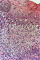
Fig.5-A2
Chronic mucocutaneous Leishmaniasis
Histologically a non-specific inflammation of the oral mucosa is seen with the presence of leishmanias laying within some vacuolated histiocytes. With this magnification and in the HE stain the few leishmanias are seen only with difficulties.
|
|

Fig.5-B2
Carcinoma
The histological examination of the biopsy shows a cornified squamous cell carcinoma of the oral mucosa.
|