|
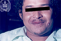
Fig.4-A1
Mucocutaneous Leishmaniasis
A 26-years old man from the State of Barinas / Venezuela developed during several weeks a skin ulcer at the left cheek. It had a diameter of 2 cm and was situated below the mouth growing steadily and was itching. Two weeks previously appeared another smaller ulcer on the right upper lip with a diameter of 1 cm.
|
|
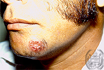
Fig.4-B1
Dermatophytosis
On the chin below the left labial angle a skin ulcer is seen with a diameter of 4 cm. In the centre a crusty material is present. The farmer comes from the same area as the patient of Fig 4-A1 and 4-A2 and his ulcer developed 2 months ago. Now it is very itching and reaches this size.
|
|
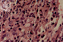
Fig.4-A2
Mucocutaneous Leishmaniasis
The histology shows a non-specific granulation tissue with several histiocytes presenting a vacuolated cytoplasm. In some of these cavities only sporadic leishmanias are seen. They are Grocott negative.
|
|
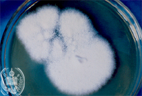
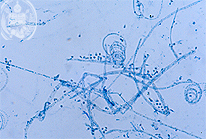
Fig.4-B2
Dermatophytosis
After finding some hyphae in the ulcer just described a culture was done which now shows a whitish colony after 20 days.
|