|
II.
Paracoccidioidomycosis
|
|
Case 16:
Paracoccidioidomycosis / Non-specific dental fistula
|
|
|
|
Paracoccidioidomycosis
|
|
Non-specific dental fistula
|
|
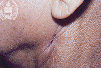
Fig.16-A1
Paracoccidioidomycosis
A 61-year old coffee farmer from the State Barinas / Venezuela observed a small fistula developing since five months in the skin of his left mandibular region below the earlobe with a discrete scarring around the fistula. In addition, he lost weight and was coughing permanently.
|
|
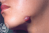
Fig.16-B1
Non-specific dental fistula
A 31-years old patient presented a small tumor-like swelling with a minimal fistula in his left mandibular region starting one year ago after the extirpation of a tooth.
|
|
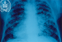
Fig.16-A2
Paracoccidioidomycosis
The x-ray examination of the thorax revealed focal opacities of different sizes in the middle fields of both lungs.
|
|
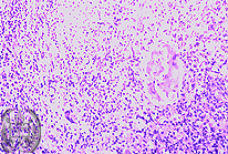
Fig.16-B2
Non-specific dental fistula
The biopsy shows a non-specific granulation tissue with the presence of some giant cells. In addition, a small necrotic bone fragment was found, apparently representing the remnant of a tooth. HE stain, low power magnification.
|
|

Fig.16-A3
Paracoccidioidomycosis
In smears from the fistula some tissue and inflammatory cells and a giant cell were found, the latter containing several relatively large yeast cells.
|
|
|
|
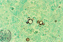
Fig.16-A4
Paracoccidioidomycosis
In the sputum of the patient in further examinations large yeast cells of the dimorphic Paracoccidioides brasiliensis with the typical multiple budding were confirmed with the Grocott method.
|
|
|
| español | english | deutsch |
|
|