|
III.
Blastomycocis
|
|
Case 25:
Blastomycocis / Carcinoma
|
|
|
|
Blastomycocis
|
|
Carcinoma
|
|

Fig.25-A1
Blastomycosis
In this patient from Cincinnati/ Ohio, USA an extensive tumor-like ulceration of the lower lip is seen extending to the chin and cheek. This is a secondary lesion after a hematogenous spread from a primary infection of the lungs caused by inhalation of fungus cells.
|
|

Fig.25-B1
Carcinoma
The tumor of the lower lip with nodular proliferations resembles the lesion of the patient described in Fig 25-A1.
|
|
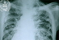
Fig.25-A2
Blastomycosis
The x-ray picture of the lungs in this case shows dense bilateral infiltrates. The apical parts of both lungs mostly do not show lesions in fungal infections, contrary to infections in the tuberculosis.
|
|
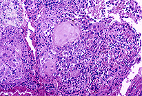
Fig.25-B2
Carcinoma
Histology reveals a squamous cell carcinoma with a slight cornification.
|
|

Fig.25-A3
Blastomycosis
The tissue reaction in cases of the infection with Blastomyces dermatitidis is usually mixed. A non-specific granulation tissue is found, frequently accompanied by giant cells and a granulomatous inflammation. Fungi are not seen clearly in this HE stain and at this low power.
|
|
|
| español | english | deutsch |
|
|