|
III.
Blastomycocis
|
|
Case 26:
Blastomycocis / Carcinoma
|
|
|
|
Blastomycocis
|
|
Carcinoma
|
|

Fig.26-A1
Blastomycosis
In the oral cavity of a patient from the USA a tumor-like proliferation with ulcerations is found. The blastomycosis was named earlier North-american (deep) mycosis. It occurs mainly in the Eastern parts of the States. Nowadays also autochthonous infections were described also in Africa and a few single cases in the Middle East.
|
|
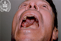
Fig.26-B1
Carcinoma
It is localized at the tongue and shows necrotic regions.
|
|
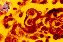
Fig.26-A2
Blastomycosis
This picture shows in the HE stain and at high power a typical solitary budding which represents the multiplication of Blastomyces dermatitidis in the tissue. The daughter cell in this is not smaller than the mother cell and sits on her with a broad base.
|
|
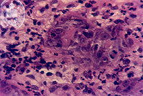
Fig.26-B2
Carcinoma
Histologically it is a squamous cell carcinoma.
|
|
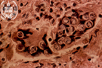
Fig.26-A3
Blastomycosis
Frequently the yeast-like cells of B. dermatitidis are detected within giant cells. This is a routine stain at low power.
|
|
|
| español | english | deutsch |
|
|