|
III.
Blastomycocis
|
|
Case 27:
Blastomycocis / Basalioma
|
|
|
|
Blastomycocis
|
|
Basalioma
|
|
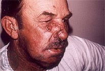
Fig.27-A1
Blastomycosis
A patient from Cincinnati / Ohio, USA shows a tumor-like proliferation under his right eye extending to the nose.
|
|
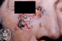
Fig.27-B1
Basalioma
This ulcerated tumor is situated beneath the eye and extends to the cheek.
|
|
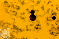
Fig.27-A2
Blastomycosis
The solitary budding of fungns cells in fissues in this case of Blastomycosis occurs with the daughter cell considerably smaller than the mother cell. Here be see a special coloration of fungus cells by the Grocott method. The multiple budding is typical of the fungus cell of P. brasiliensis
|
|
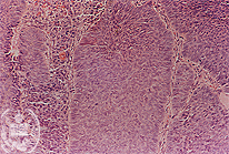
Fig.27-B2
Basalioma
Histologically this tumor is not malignant.
|
|
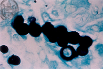
Fig.27-A3
Blastomycosis
The fungus cells of B. dermatitidis may be disposed chain-like in tissues similar to cells of Loboa loboi. Also are found marked deformations of fungus cells.
|
|
|
| español | english | deutsch |
|
|