|
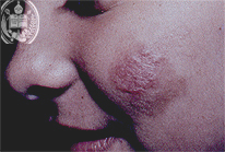
Fig.39-A1
Tuberculoid Lepra
A 13-years old schoolgirl from the town Barinas / Venezuela had a two years lasting erythematous -papilomatous skin lesion on her left cheek despite of several treatments improvement was not observed.
|
|

Fig.39-B1
Dermatophytosis
A 38-years old female patient presents on her right cheek a circular, reddish focus with a marked formation of crusts which had started several weeks ago. A biopsy was refused.
|
|

Fig.39-A2
Tuberculoid Lepra
In a small biopsy a granuloma is seen under higher power consisting of epitheloid cells. Both, the ZN and the Grocott method were negative. The Mitsuda reaction of the skin was weakly positive.
|
|
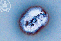
Fig.39-B2
Dermatophytosis
The culture of scraped clinical material showed after three weeks growing on Sabouraud-Glucose-Agar a whitish flocky colony with a reddish-brown border.
|
