|

Fig.48-A1
Histoplasmosis
A 61-years old female patient from the State Barinas / Venezuela presents a circular, coin-like ulceration on the left cheek. It was neither painful nor itching and had a diameter of 3 cm. clinically and histologically a Leishmaniasis was assumed. Several intracellular small granules were seen. Later, it could be confirmed that instead of a Leishmaniasis a histoplasmosis existed which could be proved by the Grocott method and by culture.
|
|

Fig.48-B1
Epithelioma
A 20-years old man from the town of Barinas / Venezuela presented on his left cheek an oval, flat tumor with an ulcer, 2,5 cm. in diameter. The regional lymph nodes were neither enlarged, nor painful. After taking a biopsy the tumor was completely removed by surgery. The further course of the disease is not known.
|
|
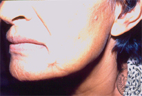
Fig.48-A2
Histoplasmosis
After 3 months of a specific treatment the ulcer on the cheek was completely cured (48-A1), but the patient lost permanently weight and died of a generalized fungus infection with Histoplasma capsulatum var. capsulatum.
|
|
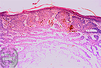
Fig.48-B2
Epithelioma
A keratotic basal cell epithelioma was dignosed in the biopsy with the formation of horny, partly calcified cysts. The tumor was resected completely.
|

