|
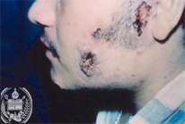
Fig.19-A1
Paracoccidioidomycosis
In a 33-years old farmer from the Venezuelan Andes near Barinas several fast growing ulcers of the skin on the left cheek had developed. On the left side of the jaw some lymph nodes were palpable. His general state of health was good; only occasionally he was short-winded.
|
|
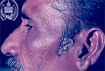
Fig.19-B1
Naevus cell naevus
A 62 -years old farmer from the same region as the patient described in-A1 presents an ovally shaped great pigmented skin ulcer covered by crusts in his left auricular region. Lymph nodes were not palpable. The patient is in a good state of health.
|
|
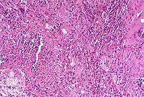
Fig.19-A2
Paracoccidioidomycosis
The biopsy taken from a skin ulcer shows a so called "mixed cell reaction" with granulation tissue and some giant cells. The large vacuolic yeast -like cells of P. brasiliensis are hardly to see in this low power and with the HE stain.
|
|
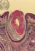
Fig.19-B2
Naevus cell naevus
The histological examination of the biopsy taken from the border of the ulcer reveals hyperkeratoris follicular and nests of pigmented naevus cells without any signs of malignancy. The complete naevus with the ulcer could be removed surgically.
|