|
II.
Paracoccidioidomycosis
|
|
Case 20:
Paracoccidioidomycosis / Melanoma
|
|
|
|
Paracoccidioidomycosis
|
|
Melanoma
|
|
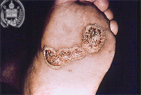
Fig.20-A1
Paracoccidioidomycosis
A merchant from Mérida / Venezuela noted a tumor-like, ulcerated skin lesion at his left plantar surface which had grown slowly during some months. The doctor who was consulted made a smear from the ulcer surface without staining it. The result will be shown in Fig. 20-A2.
|
|
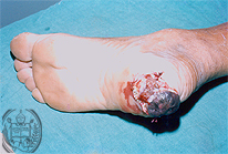
Fig.20-B1
Melanoma
A farmer living near the town of Mérida in Venezuela noted a slowly growing tumor-like swelling in the posterior parts of his left sole. The skin of this tumor was partly blackish and partly ulcerated.
|
|
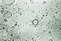
Fig.20-A2
Paracoccidioidomycosis
In this smear a structure was found which resembled the steering wheel form of a budding Paracoccidioides brasiliensis cell, however, it could represent air bubbles.
|
|
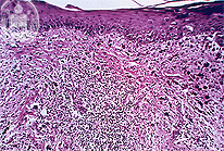
Fig.20-B2
Melanoma
Histology revealed a malignant tumor of this kind.
|
|
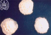
Fig.20-A3
Paracoccidioidomycosis
In the culture of the material taken from the ulcer surface did grow colonies of Paracoccidioides brasiliensis.
|
|
|
|
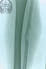
Fig.20-A4
Paracoccidioidomycosis
The patient suddenly accused pain at his left lower extremity. In the x-ray examination of the fibula a localized defect was detected which represented a osteomyelitis caused by this fungus. This confirmed a marked haematogenous dissemination of P. brasiliensis.
|
|
|
|
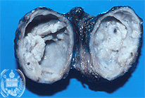
Fig.20-A5
Paracoccidioidomycosis
In an enlarged axillary lymph node also was found a fungal infection with P. brasiliensis which had produced necrotic and caseating foci.
|
|
|
| español | english | deutsch |
|
|