|
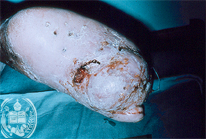
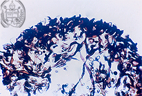
Fig.55-A1
Eumycetoma
A patient from Mérida / Venezuela discovered on the heel of his foot a pooly limited tumorous nodule with a localized hemorrhage. Before we describe the other details of the mycetomas we would like to give a definition of this disease. Its name earlier was used only for fungal infections, but nowadays after a reclassification the name is applied for bacterial infections also while the name mycetoma is maintained. They occur in three types and this tumor-like lesion is situated exclusively in the skin, eventually with formation of fistulae. In the abscesses present in the dermis are found the so called grains (accumulations of germs).
|
|
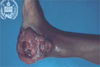
Fig.55-B1
Fibrosarcoma protuberans
A patient from the Venezuelan Andes presents in the posterior part of his plantar surface a large, well limited tumor.
|
|
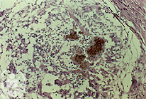
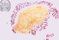
Fig.55-A2
Eumycetoma
Histologically the abscesses in the inflammatory tissue of the corium show grains of different sizes and forms, stained with haematoxylin. In the eumycetomas the grains consist of accumulations of true fungi.
|
|
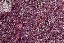
Fig.55-B2
Fibrosarcoma protuberans
The histology confirms the clinical diagnosis.
|